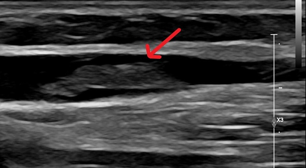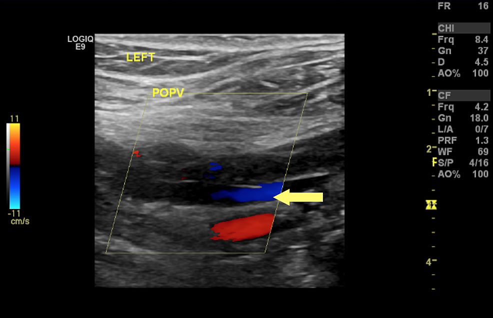

Chronic Venous Insufficiency ExamĬVI STUDY. If present, these patients need to be treated with blood thinners to reduce the risk of further complications. Patients with leg (or arm) swelling can be evaluated with a venous ultrasound exam for the presence of a blood clot, often called a " DVT" (deep vein thrombosis).
hepatic veno -occlusive disease (e.g.VENOUS DOPPLER is a specialized ultrasound procedure most commonly performed in the legs but sometimes in the arms or neck. often associated with spectral broadening of the Doppler envelope and truncation of the a wave.  decrease in phasicity (diminution of anterograde/retrograde velocities). this presumes tricuspid valvular competence. maintenance of a dominant anterograde S wave > D wave. abnormally high amplitude of the a and v waves. increased pulsatility (exaggeration of anterograde/retrograde velocities). The consequent hemodynamic perturbations may manifest as: Differential diagnosisĪlterations in the normal hepatic venous Doppler waveform often indicate cardiac dysfunction, although it may also reflect disease of the hepatic parenchyma and/or vasculature. Sometimes a c wave occurs as a second small inflection above the baseline, right after the a wave, reflecting the effect of the tricuspid valve bulging into the right atrium. this wave is almost always lower in magnitude than the s wave. as the tricuspid valve opens, blood flows from the right atrium into the right ventricle, resulting in a net flow of blood away from the liver and the waveform again dives back down below the baseline. if the atrium fills to capacity then there may be a small amount of flow "recoil" backward, resulting in a v wave that rises above the baseline. as blood fills the right atrium, the flow from the hepatic veins and IVC slows, resulting in the s wave returning back to baseline. this typically forms the highest velocity deflection seen in the waveform. the resultant "stretching" of the right atrium results in a drop in pressure, creating a gradient for anterograde flow from the inferior vena cava and hepatic veins, most pronounced at mid-systole. as systole commences, right ventricle contraction results in longitudinal, apically oriented traction on the tricuspid annulus. the small reversal of flow typically results in a small wave above the baseline, reversed from the overall net flow back to the heart.
  this also creates a pressure gradient favoring a lesser degree of retrograde flow into the IVC and hepatic veins. coinciding with the "p wave" on the electrocardiogram, contraction elevates pressure within the right atrium creating a gradient for late diastolic filling of the right ventricle.
The normal periodic hepatic vein waveform is typically described in four parts: Some prefer the term "periodic" since the term "triphasic" already has a specific application in arterial spectral Doppler waveforms and since "periodic" suggests that the waveform is transmitted by cardiac motion rather than systolic flow. Multiple terms have been used to describe the hepatic vein waveform, including "phasic", "triphasic", "tetrainflectional", and "periodic". The shape of the hepatic vein spectral Doppler waveform is primarily determined by pressure changes in the right atrium, or more exactly the blood flow resulting from the resultant pressure gradients.


 0 kommentar(er)
